The current direction of the medical imaging market is to provide patients with more comfortable and safe solutions. According to the recently released Frost & Sullivan[i] report, the current trends in medical imaging have the following:
· Reduce and manage ionizing radiation doses and exposures;
· Mobile, portable, wireless solutions;
· Elastography techniques for assessing tissue stiffness;
· Dedicated to developing technologies that improve patient comfort and experience;
· Entry level advanced imaging equipment.
Ultrasound can achieve all of the above features. However, in order for ultrasound examination to be more widely used and user-friendly, image quality must be improved. With better image quality, faster, more accurate diagnostics can be performed without compromising patient safety and comfort.
Ultrasound equipment can be used for all types of examinations, whether obstetric and gynecological imaging, or cardiac or radiological imaging. As ultrasound systems become more advanced and complete, users are becoming more professional and skilled in using these systems.
The miniaturization of ultrasound equipment helps to expand the range of bedside applications, especially when clinicians use ultrasound equipment to diagnose at the bedside. Because ultrasound requires more practical and specialized operations by the user than other methods of examination, experience is essential in ultrasound. If the doctor is unable to obtain the image required for the diagnosis, the patient will be transferred for additional MRI or CT examination. This not only wastes time and resources, but also exposes patients to unnecessary radiation doses during CT examinations, causing safety problems.
Ultrasound technology development roadmap
With the continued development of this trend and the expansion of the ultrasound market in new applications, new methods of ultrasound use are emerging. Ultrasound technology can now be used in cardiology, breast biopsy, gastroenterology, head and neck surgery, neonatal, neurology, ophthalmology, vascular, pulmonary disease, urology, musculoskeletal, emergency, and Other areas.
For bedside testing, clinicians want the best image quality with just one button. This requires that the ultrasound system must be more intuitive and independent of the operator, allowing doctors with less ultrasound experience to get the best image quality faster and easier.
Although ultrasound systems are developing rapidly and more effectively, the industry is still facing a shortage of ultrasound experts. In order to make ultrasound examinations more widely available and easier to use, the importance of image quality cannot be ignored.
In addition, ultrasound systems require multiple uses in a shared service environment. In this environment, different clinical applications have different image quality requirements. Ultrasound systems must be flexible, versatile, and easy to use to improve diagnostic precision and scan time. This specifies the image quality to ensure accurate, faster diagnosis for each clinical application supported by the system.
Improve image quality for more accurate diagnosis
Image quality in ultrasound images is critical. For good image quality, you need a good image enhancement device. Due to the increasingly fierce competition among equipment manufacturers, there are many different types of processing methods available, and these processing methods have different adaptability to actual image signals. When the image enhancement processing method is not adaptive enough, the image may lose important information, and artifacts may appear in the image. The adaptive filter does not produce image processing artifacts, which allows the image to truly reflect the physical signal content.
For clinicians, image quality enhancement refers to the appearance of small noise, clear lines, and good visualization of organ boundaries and textures. For shared services, image quality must not only adapt to image signals, but also to clinical applications to meet the needs of clinicians and experts. For bedside detection systems, images should be easy to interpret, even for clinicians with limited ultrasound experience. Real-time image processing is essential because multiple diagnostics may be required during the inspection process. For all clinical applications, the results should be accurate diagnosis and rapid delivery.
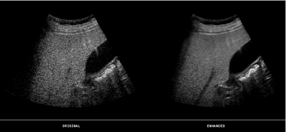
Note: The left side is the unprocessed liver image and the right side is the processed liver image. High-resolution images, clear blood vessels, and processed images remove spots and achieve superior edge sharpening and contrast enhancement.
Automatic measurement technology
Automatic measurement is another aspect that requires high image quality. Automated measurement of ultrasonography for intima-media thickness (IMT) for the detection of atherosclerotic disease in humans and may also be used to track the regression, containment or progression of atherosclerosis.
Manual measurements are not only time consuming, but the results depend on training and subjective judgment. Automated measurement results are less dependent on the reader's experience, and the results of automatic measurements by the user are more stable than older manual analysis systems.
Automated measurements are more efficient and can be used to automatically detect the internal and external skull boundaries of the fetal head in an ultrasound image. These boundaries are used to measure the double top diameter (BPD) and head circumference (HC). In the urology department, the autonomic measurement technique is used to measure the bladder weight. Ultrasound measurement of bladder weight (UEBW) is an important diagnostic indicator of bladder outlet obstruction.
These measurements require images with clear boundaries and line connections and high contrast to allow measurements to be made with high accuracy.
3D/4D inspection
As 3D/4D ultrasound technology becomes more and more personalized, inspections are becoming more and more valuable, and even provide more accurate diagnostic results. At present, the system can not only provide fetal facial images for obstetrics - it can provide more clinical value by finding brain abnormalities, detecting cleft palate, finding and locating tumors in general images, and also can be used for ejection fraction and regional dyskinesia. / Exercise can't perform an accurate heart check and visualize the heart valve.
3D ultrasound examinations provide a wealth of information. To generate good image quality that matches this large amount of information, image enhancement processing techniques must be very advanced and complete, especially when used in 4D (ie, under real-time conditions). Adaptive processing techniques will analyze each voxel to distinguish between real information and artifacts, such as noise and speckle. Real information can also be enhanced to make the visualization of boundaries and structures more precise. Processing all of this information in real-time conditions requires very advanced software and hardware.
According to Thomas Jefferson University (comparison of image processing techniques for improving 3D ultrasound images, Flemming Forsberg et al. 2010): "The new three-dimensional ultrasound image enhancement technology has been evaluated in abdominal and obstetric applications. The voxels and voxels processed with 2D image enhancement software, the new 3D processing technology performs best."
3D/4D examination also plays an important role in breast examination. There have been some recent discussions on whether women with high breast tissue density should undergo mammography (x-ray) examination. This shows that ultrasound technology plays a very important role in health care. Now that breast volume measurement is feasible, ultrasound technology can cope with the difficulties faced by mammography. The location of the tumor, as well as the malignant and benign conditions, are clearly shown and, most importantly, less than 15 minutes from the start of the examination to the diagnosis.
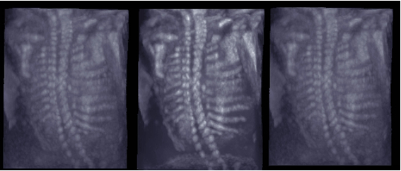
Note: Fetal spine images acquired by 3D technology, rendered. On the left is the original image of the untreated spine, with a 3D enhanced spine image in the middle and a 2D enhanced spine image on the right. Note the sharp outline of the fetal spine in the 3D enhanced image.
Patient's comprehensive safety
Adequate 2D/3D/4D image processing technology ensures patient safety when using a variety of different ultrasound machines, bedside detection, shared services and very advanced equipment.
The development of 3D/4D technology will reduce the chances of other (checking) methods involving ionizing radiation. This is the future of the ultrasound market – in all applications, 3D/4D can display more realistic images, providing the best image quality for less experienced doctors, helping them to make accurate diagnoses immediately. With this technology, patients or clinicians are not exposed to radiation, and the use of mobile devices can provide patients with a more comfortable testing experience.
About the author 
Isabelle Wegmann Hachette is the Senior Application Manager of TECVision, a leading developer of strategic products, undertaking technical management of major customers and solving installation problems for customers in many regions. She holds a Master of Science in Electrical Engineering from the Image Processing Laboratory at Linköping University in Sweden and has also served as a lecturer at the University.
AC Contactor switch mainly used for making or breaking circuit at a long distance, suitable for controlling starting\stopping\reversing of AC motor. AC Contactor of korlen conforms to the requirement of IEC60947-4-1& GB14048.4 standards.
Except AC Contactor, there are many different types of low voltage electric appliances, such as Thermal Relay,Manual Motor Startor ,led light, Circuit Breaker, etc.
AC Contactor
AC Contactor,Magnetic Contactor,Wafer Style Valve
Wenzhou Korlen Electric Appliances Co., Ltd. , https://www.korlenelectric.com